Last 5 Year PYQs in Ophthalmology for NEET PG
Jan 10, 2025

Do you also among the thousands of other NEET PG aspirants feel that Ophthalmology is a tricky subject to master? With so many clinical scenarios, overlapping conditions, and tricky terminologies, you feeling overwhelmed is natural. However there is one way that can make all the difference
Analyzing previous years’ questions can help you spot patterns, understand high-yield topics, and give you a real sense of what to expect in the exam.
In this blog, we’ve compiled some of the most important Ophthalmology questions from the last five years. And, we’ve also given the answers and explanations. So, let’s dive in and decode the most frequently asked concepts in Ophthalmology.
Download FREE PDFs of Last 5-Year NEET PG PYQs – All Subjects
Q1. Which layer is accountable for preserving the moisture and clarity of the cornea?
- Descemet’s membrane
- Endothelium
- Stroma
- Corneal epithelium
Ans. 1) Endothelium
- The corneal endothelium is primarily responsible for maintaining the hydration and transparency of the cornea
Q2. What is the distinguishing characteristic of a fungal ulcer?
- Hypopyon
- Dendritic ulcer on a fluorescein dye
- Ring abscess
- Satellite lesion
Ans. 4) Satellite lesion
- Fungal infection typically presents with a dry /rough central corneal ulcer with feathery edges (hyphae) and it may present as a satellite (single) lesion that is infective and immune-reactive.
- These satellite lesions are often seen as smaller infiltrates or white fluffy patches adjacent to the primary ulcer.
Also read: Last 5 Years PYQs in Radiology for NEET PG
Q3. What is the term for the additional row of eyelashes located behind the grey line?
- Trichiasis
- Distichiasis
- Tylosis
- Madarosis
Ans. 2) Distichiasis
- Distichiasis is a condition in which the eyelashes grow abnormally and emerge from the Meibomian gland openings behind the normal lash line.
- These extra lashes can cause irritation and damage to the cornea and conjunctiva, leading to symptoms such as eye discomfort, redness, tearing, and sensitivity to light.
- Treatment options for distichiasis include manual plucking of the extra lashes, cryotherapy (freezing the hair follicles), or various surgical procedures to remove or redirect the aberrant eyelashes.
Q4. A middle-aged woman presented with bilateral proptosis, restriction of eye movements, and chemosis. Her thyroid profile showed she was hyperthyroid. What is the most probable cause?
- Orbital pseudotumor
- Grave’s ophthalmopathy
- Orbital lymphoma
- Orbital cellulitis
Ans. 2) Grave’s ophthalmopathy
Grave’s ophthalmopathy, also known as thyroid-associated ophthalmopathy, is an autoimmune condition commonly associated with Graves' disease, which is a form of hyperthyroidism.
- It is characterized by inflammation and swelling of the tissues surrounding the eyes, including the muscles and fatty tissues within the orbit.
- The condition often presents with the following features:
- It is the most common cause of proptosis: unilateral/bilateral, painful/non-painful, axial/non-axial.
- Grittiness and red eyes.
- Lid retraction.
Q5. The purpose of the photostress test is to distinguish between
- Macula and optic nerve disease
- Cataract and glaucoma
- Lens and cornea
- Retina and vitreous pathologies
Ans. 1) Macula and optic nerve disease
- Photostress Test helps in this differentiation:
- Macular Diseases: If the macula is affected by conditions such as age-related macular degeneration (AMD), macular edema, or macular dystrophies, the recovery time after the photostress exposure may be significantly prolonged around 60 sec.
- Optic Nerve Diseases: Optic nerve diseases, such as optic neuritis, optic nerve ischemia, or optic nerve degeneration. In these conditions, the macula is relatively spared, and the recovery time after the photostress exposure remains within the normal range, i.e., around 30 to 50 seconds.
Also read: Last 5 Year PYQs in Pharmacology for NEET PG
Q6. Shifting fluid sign is seen in?
- Exudative retinal detachment
- Traction retinal detachment
- Rhegmatogenous retinal detachment
- Retinal dialysis
Ans. 1) Exudative retinal detachment
- The shifting fluid sign is typically seen in exudative retinal detachment (RD).
- Exudative retinal detachment is a condition in which fluid accumulates beneath the retina, leading to its separation from the underlying layers. The shifting fluid sign refers to the movement or shifting of subretinal fluid with changes in body or head position.
Q7. In a 40-year-old patient with diabetes, a squint was observed. The secondary deviation was higher than the primary deviation during examination. The forced duction test yielded negative results. Which type of squint is expected to be predominant in this individual?
- Comitant squint
- Restrictive squint
- Paralytic squint
- Pseudosquint
Ans. 3) Paralytic squint
- In the case of a 40-year-old patient with diabetes who presents with a squint, where the secondary deviation is higher than the primary deviation and the forced duction test yields negative results, this is suggestive of paralytic squint.
- Paralytic squint causes greater deviation in the yoke muscle of the sound eye compared to the eye with the paralyzed muscle. This is due to the strong impulse of innervation required to fix the paralyzed eye, which is transmitted to the yoke muscle of the sound eye. This is based on Hering's law of equal innervation.
- A forced duction test is done to differentiate the mechanical causes from extraocular muscle palsy causes of incomitant squint. It is negative in incomitant squints caused by extraocular muscle palsy (paralytic squint).
Also read: Last 5 Year PYQs in Forensic Medicine
Q8. Which of the following conditions is associated with the findings observed by a physician during an eye examination of a small child?

- Myopia
- Astigmatism
- Hyperopia
- Emmetropia
Ans. 3) Hyperopia
- The image is suggestive of convergent squint.
- High hyperopia/hypermetropia (+4 to +7D) results in increased exertion of accommodation and results in convergent squint.
Q9. Sympathetic ophthalmia is seen after _____?
- Penetrating trauma
- Blunt trauma
- Chemical injury
- Ocular infection
Ans. 1) Penetrating trauma
- Sympathetic ophthalmia is most commonly observed as a complication following penetrating ocular trauma, in which intraocular antigens are exposed to the immune system, triggering an autoimmune response that affects both eyes.
Q10. What is the likely reason for a patient showing both unilateral and bilateral abducent palsy?
- Retinoblastoma
- Orbital pseudotumor
- Orbital cellulitis
- Cavernous sinus thrombosis
Ans. 4) Cavernous sinus thrombosis
- Cavernous sinus thrombosis is the most probable cause of both unilateral and bilateral abducens (abducent) palsy. Cavernous sinus thrombosis occurs when a blood clot forms within the cavernous sinus, a venous sinus located at the base of the skull behind the eyes. The cavernous sinus contains the cranial nerves, including the abducens nerve (cranial nerve VI), which innervates the lateral rectus muscle responsible for abduction (outward movement) of the eye.
Also read: Last 5 year PYQs in Microbiology for NEET PG
Q11. Where is the location of the lesion in Wernicke's hemianopic pupil?
- Optic nerve
- Optic tract
- Optic radiation
- Optic chiasma
Ans. 2) Optic tract
- In Wernicke's hemianopic pupil, the site of the lesion is typically in the optic tract.
- The optic tracts contain both visual and pupillomotor fibers. The visual fibers terminate in the lateral geniculate body, and pupillary fibers leave the optic tract and terminate in the pretectal nuclei.
- Wernicke hemianopic pupil is characterized by normal pupillary light reflex when the unaffected hemiretina is stimulated and absent when the involved hemiretina is stimulated.
Q12. A contact lens user presents with the following clinical picture. He has had watering, redness, and foreign body sensation in the eye for the past 2 months. What is the most probable diagnosis?

- Trachoma
- Follicular conjunctivitis
- Giant papillary conjunctivitis
- Spring catarrh
Ans. 3) Giant papillary conjunctivitis
- The clinical presentation of watering, redness, and foreign body sensation, along with an image showing large papillae in the upper tarsal conjunctiva in a contact lens wearer, is suggestive of giant papillary conjunctivitis.
- Giant Papillary Conjunctivitis (GPC), aka mechanically-induced papillary conjunctivitis, is a localised allergic response to a physically rough or deposited surface (contact lens, prosthesis, nylon suture, and scleral buckle).
Also read: Last 5 Years PYQ's in Anesthesia for NEET PG
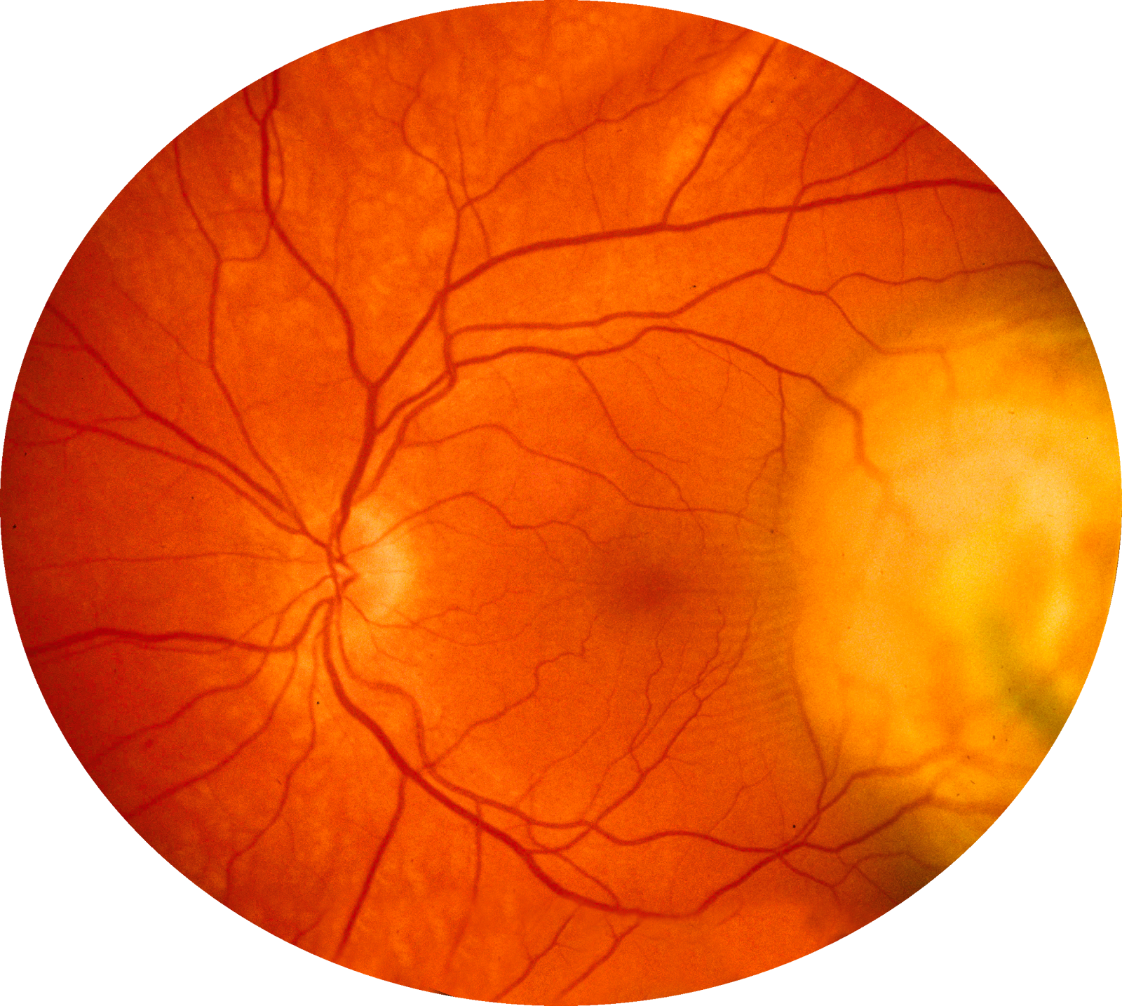
Q13. A 60-year-old patient with progressive painless loss of vision presents to the ophthalmology OPD. The fundus image of the patient is given below. What could be the probable finding and the cause of it?

- Soft exudate, hypertension
- Hard exudate, diabetes mellitus
- Flame-shaped hemorrhages, hypertension
- Soft exudate, central retinal vein occlusion
Ans. 2) Hard exudate, diabetes mellitus
- Based on the provided fundus image showing hard exudates in the retina, the most probable finding and cause could be diabetes mellitus.
- Hard exudates are yellowish deposits in the retina that result from the leakage of lipids and proteins from damaged blood vessels. They are typically seen in conditions involving retinal vascular changes.
Q14. What would be the suitable course of action for managing a 15-year-old girl who is non-adherent to wearing spectacles for her myopic astigmatism?
- LASIK
- Spherical equivalent spectacles
- Femto LASIK
- Implantable collamer lens
Ans. 2) Spherical equivalent spectacles
- The most appropriate management for a 15-year-old girl who is not compliant with spectacles for her myopic astigmatism would be spherical equivalent spectacles
- Spherical equivalent spectacles provide correction for both myopia (nearsightedness) and astigmatism. They are a non-invasive and reversible option that can effectively improve visual acuity and provide clear vision when worn.
- In the case of a non-compliant patient, it is important to choose a management option that is simple, convenient, and easy to use.
- Spherical equivalent spectacles meet these criteria as they only require the patient to wear the prescribed spectacles during waking hours to achieve clear vision.
- Spectacles are readily available, easily adjustable, and can be customized to the patient's specific refractive error.
Also read: INI-CET Previous Year Question Papers
Q15. The person in the image is at risk of developing?

- Exposure keratopathy
- Restricted eye movements
- Diplopia
- Amblyopia
Ans. 1) Exposure keratopathy
- The person is at risk of developing Exposure keratopathy is a condition where the cornea becomes dry and damaged due to inadequate eyelid closure or incomplete blinking, leading to exposure of the cornea to the environment.
- It manifests as irritation, pain, foreign body sensation, and blurred vision.
- In severe cases, it can cause corneal ulceration and scarring.
Q16. What is the most probable diagnosis for a 7-year-old patient who presented with a white pupillary reflex and underwent enucleation, with histopathological examination revealing the presence of Flexner-Winterstein rosettes?
- Retinoblastoma
- Medulloblastoma
- Rhabdomyosarcoma
- Astrocytoma
Ans. 1) Retinoblastoma
- The most likely diagnosis based on the histopathological examination showing Flexner-Winterstein rosettes is retinoblastoma.
Microscopic pathology of Retinoblastoma
- It is divided into differentiated and undifferentiated retinoblastoma.
- The differentiated type has three forms
- Flexner-Wintersteiner Rosette are the columnar cells with a central lumen
- The rosette is called the Homer-Wright rosette
- Fleurettes
Q17. What is the diagnosis?

- Intraocular foreign body
- Pseudoexfoliation syndrome
- Ocular trauma
- Vossius ring
Ans. 2) Pseudoexfoliation syndrome
- The image reveals gray-white fibrillary material on the anterior surface of the lens derived from extracellular matrix metabolism is suggestive of pseudoexfoliation syndrome.
- Pseudoexfoliation syndrome is characterized by the deposition of fibrillar material on various ocular structures, including the lens capsule, zonular fibers, iris, trabecular, and conjunctiva.
Also read: NEET PG Previous Year Question Papers of Last 6 Years
Q18. What will be observed in a patient diagnosed with sympathetic ophthalmitis and a history of penetrating eye injury?
- Acute anterior uveitis
- Pars planitis
- Panuveitis
- Chronic anterior uveitis
Ans. 3) Panuveitis
- Sympathetic ophthalmitis is a bilateral granulomatous panuveitis occurring after penetrating trauma to one eye.
- Panuveitis involves all three uveal tissues: the iris, ciliary body, and choroid.
- The primary site of inflammation are
- Anterior chamber
- Vitreous
- Retina
- Choroid
Q19. What should be the subsequent course of action in managing a diabetic patient who has a visual acuity of 6/9 in one eye and further investigations reveal preretinal hemorrhages with neovascularization at the optic disc?
- Focal laser photocoagulation
- Pan-retinal photocoagulation
- Grid laser photocoagulation
- Scleral buckling
Ans. 2) Pan-retinal photocoagulation
- Panretinal Photocoagulation (PRP) is a laser treatment and it is used in patients who have grown abnormal new blood vessels in the drainage system of the eyeball or in the retina at the back of the eye.
- The PRP laser treatment contracts the existing blood vessels while inhibiting the growth of abnormal new blood vessels on the retina and in the drainage system of the eyeball.
Q20. What would be the recommended treatment for simple myopic astigmatism among the options provided?
- +1.00 DS
- -1.00 DC × 180 Degree
- -1.00 DS
- -1.00DS – 1.00 DC × 180 Degree
Ans. 2) 1.00 DC × 180 Degree
- Simple myopic astigmatism refers to a condition where there is myopia (nearsightedness) along with astigmatism in one eye and normal other eye.
- This prescription that indicates a correction for myopic astigmatism is -1.00 DC × 180 Degree.
- The -1.00 DC (D- dioptre and C- cylindrical) represents the cylindrical power, which addresses astigmatism. A negative cylindrical power indicates that the astigmatism is in the myopic (nearsighted) direction.
- The "× 180 Degree" denotes the axis of astigmatism. In this case, the astigmatism is at 180 degrees, meaning that the irregular curvature of the cornea or lens causing the astigmatism is aligned vertically.
- By prescribing -1.00 DC × 180 Degree, the correction aims to address both the nearsightedness (myopia) and the astigmatism in the correct axis. This combination is appropriate for a patient with simple myopic astigmatism.
Also read: NEET PG/FMGE Ophthalmology Important Questions With Answers
Download the PrepLadder app now and unlock a 24-hour FREE trial of premium high-yield content. Access Video Lectures, digital notes, QBank, and Mock Tests for best neet pg ophthalmology online coaching. Elevate your study experience and gear up for success. Start your journey with PrepLadder today!

PrepLadder Medical
Get access to all the essential resources required to ace your medical exam Preparation. Stay updated with the latest news and developments in the medical exam, improve your Medical Exam preparation, and turn your dreams into a reality!
Top searching words
The most popular search terms used by aspirants
- NEET PG Ophthalmology
- NEET PG Ophthalmology Preparation
- NEET PG Previous Year Question Paper
PrepLadder Version X for NEET PG
Avail 24-Hr Free Trial