Image Based Questions in Anatomy for NEET PG 2025
Apr 24, 2025

Q1. Which day during Week 1 of embryonic development matches with the given image?

- Day 1
- Day 2
- Day 4
- Day 5
Answer: 3) Day 4
Explanation:
- The first week of development begins with fertilization and ends with the initial stages of implantation.
- The image given shows morula formation, where the zygote becomes a solid ball of 16-32 cells.
- Cleavage: Once the zygote has reached the two-cell stage, it undergoes a series of mitotic divisions, which become smaller with each cleavage division known as blastomeres.
Q2. What structure forms at the surface of the epiblast to initiate gastrulation?
- 2
- 3
- 4
- 1
Answer: 3) 4
Explanation: Primitive streak/groove (structure-4) forms at the surface of the epiblast to initiate gastrulation.
Markings in image:
1 - Prechordal plate
2 - Primitive pit
3 - Primitive node
4 - Primitive streak/ groove

Q3. A stillborn baby was noted to have the following findings.

The above pathology is derived from which among the following structures shown in the image below?

- 1
- 2
- 3
- 4
Answer: 2) 2
Explanation: The pathology shown is Oropharyngeal teratoma, arises from Primordial germ cells that fail to migrate to the gonadal ridge and Primordial germ cells (PGC) in the image corresponds to “2”.
Markings in the image are:
1 - Allantois
2 - Primordial Germ cells (Option 2)
3 - Yolk sac
4 - Heart
Q4. A patient with a history of 6 weeks of amenorrhea and a positive urinary pregnancy test underwent an ultrasonography, which showed the following:
The marked structure in the image communicates via a duct with which of the following parts of the gastrointestinal system?

- Foregut
- Midgut
- Hindgut
- Cloacal membrane
Answer: 2) Midgut
Explanation:
- The marked structure in the image is the Yolk sac.
- The Midgut of the embryo communicates with the yolk sac via a stalk called the Vitelline duct.
Q5. The structure “b” fuses with structure “c” during the 3rd month of pregnancy. Identify the structure “c”.

- Decidua capsularis
- Decidua basalis
- Myometrium
- Decidua parietalis
Answer: 4) Decidua parietalis
Explanation:
The structure “c” is the decidua parietalis that fuses with the growing decidua capsularis around the embryo.
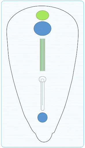
Q6. What is the structure marked “C” formed of?

- Trophoblast + Somatopleuric EEM
- Trophoblast+ Splanchnopleuric EEM
- Ectoderm + EEM
- Amniogenic cells + Somatopleuric EEM
Answer: 1) Trophoblast + Somatopleuric EEM
Explanation:
The structure marked “C” is the chorion, which is formed by the parietal (or somatopleuric) extraembryonic mesoderm and the trophoblast.

Q7. What is the type of cartilage found in the structure marked in the following radiograph?

- Hyaline cartilage
- Calcified cartilage
- Fibrocartilage
- Elastic cartilage
Answer: 3) Fibrocartilage
Explanation: The structures marked in the given radiograph is pubic symphysis and the type of cartilage is fibrocartilage.
- Fibrocartilage is a type of cartilage that is more rigid and durable compared to the other types of cartilage, such as hyaline and elastic cartilage.
- It is characterized by its dense arrangement of collagen fibers, which gives it strength and the ability to resist compressive forces.
Q8. Which of the following structural abnormalities is most commonly associated with the given image?

- Malformation of the vertebral arches
- Incomplete development of the basichondrocranium
- Herniation of brain tissue through the skull
- Fusion of the cranial sutures
Answer: 2) Incomplete development of the basichondrocranium
Explanation: Anencephaly is associated with severe abnormalities in the base of the skull (basichondrocranium), affecting its shape, position, and ossification.
Anencephaly:
- Neurulation failure: Anencephaly occurs when the neural tube fuses in the spinal cord (caudal neuropore) but fails to fuse in the brain region (cranial neuropore), leading to missing parts of the brain and skull.
- The cranial and caudal neuropores close around days 25 and 28 of development, respectively, in a normally developing fetus.
Q9. Which of the given options are correctly matched based on the image given?

- 3- Lateral recess, 5- Frontal horn
- 1- Occipital horn, 7- Third Ventricle
- 2- Fourth Ventricle, 4- Aqueduct
- 3- Temporal horn, 6- Aqueduct
Answer: 3) 2- Fourth Ventricle, 4- Aqueduct
Explanation:
- Each cerebral hemisphere contains one of the two large lateral ventricles. This ventricle is roughly C-shaped and can be divided into several sections.
- Body
- Anterior horn - extends into the frontal lobe
- Posterior horn -extends into the Occipital lobe
- Inferior horn - extends into the Temporal lobe
- The third ventricle is a narrow space located between the two thalami. It connects to the lateral ventricles in the front via the interventricular foramina (of Monro) and to the fourth ventricle in the back through the cerebral aqueduct (of Sylvius).
- The cerebral aqueduct, also known as the aqueduct of Sylvius, is a narrow passage approximately 1.8 cm long that links the third ventricle to the fourth ventricle.
- The fourth ventricle is sandwiched between the brainstem and the cerebellum.
- From the fourth ventricle, the CSF moves into the central canal of the spinal cord.

Q10. Which of the following intrinsic neurons are correctly matched with their respective layers in the cerebellar cortex?

- 1- Plexiform layers; Golgi cells
- 2- Molecular layer; Purkinje cells
- 2- Granular layer; Basket cells
- 3- Granular layer; Golgi cells
Answer: 4) 3- Granular layer; Golgi cells
Explanation:
- Golgi cells are found in the granular layer of the cerebellar cortex.
- They are inhibitory interneurons that play a role in modulating the activity of granule cells.
Q11. A 62-year-old male presents to the emergency department with a sudden onset of severe vertigo, bilateral hearing loss, and tinnitus. He also exhibits facial weakness on the right side and demonstrates ataxia when attempting to walk. An MRI performed to evaluate the condition is given below. Which artery is most likely affected in this patient’s condition?

- Middle cerebral artery
- Superior cerebellar artery
- Anterior inferior cerebellar artery
- Vertebral artery
Answer: 3) Anterior inferior cerebellar artery
Explanation:
- The symptoms—severe vertigo, bilateral hearing loss, tinnitus, and facial weakness—are indicative of an AICA stroke.
- The MRI image shows a B/L infarct in the anterior cerebellum, which further confirms the involvement of AICA.
- The AICA arises from the caudal third of the basilar artery and supplies the inner ear, lateral pons, and the anterior inferior cerebellum.
Q12. Which of the following conditions is associated with lesions at the level marked in the midbrain?

- Weber’s syndrome
- Benedikt’s syndrome
- Parinaud’s syndrome
- All of the above
Answer: 4) All of the above
Explanation:
The superior colliculus is marked in the above image and all the given syndromes are associated with lesions at this level.
Q13. Which among the following are pneumatic bones?

- 1,2,3,6,7
- 1,2,3,5,7
- 1,2,4,6
- 1,3,5,6,7
Answer: 1) 1, 2, 3, 6, 7
Explanation: Labelings in the image are:
1 - Frontal
2 - Sphenoid
3 - Maxilla
4 - Mandible
5 - Parietal
6 - Temporal
7 - Mastoid

Pneumatic bones:
- Frontal (1): Contains sinuses
- Sphenoid (2): Contains sinuses
- Maxilla (3): Contains sinuses
- Temporal (6): Contains ear cavity
- Mastoid (7): Contains mastoid air cells
Q16. Identify the marked branch of the External carotid artery?
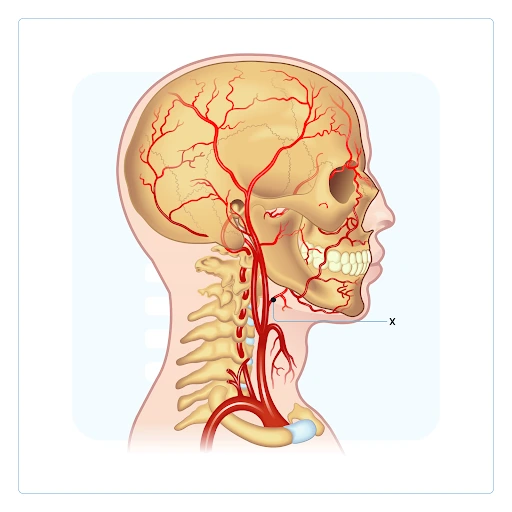
- Lingual Artery
- Facial Artery
- Maxillary Artery
- Superior Thyroid Artery
Answer: 1) Lingual Artery
Explanation:
Lingual artery:
- A major branch of the external carotid artery.
- Supplies the tongue and the oral floor.
- Arises anteromedially from the external carotid artery.
- Located at the tip of the greater horn of the hyoid bone, between the superior thyroid artery and the facial artery.
Q17. From the sagittal section of the larynx given below, identify the part marked as X.

- Thyroid cartilage
- Cricoid cartilage
- Arytenoid cartilage
- Cuneiform cartilage
Answer: 3) Arytenoid cartilage
Explanation:
- Epiglottis: Leaf-shaped cartilage located at the superior aspect of the larynx
- Thyroid cartilage: The largest cartilage of the larynx, visible as a large shield-like structure with its anterior prominence forming the Adam's apple.
- Cricoid cartilage: A ring-shaped cartilage below the thyroid cartilage, marking the lower limit of the larynx.
- Arytenoid cartilages: Pair of cartilages located on top of the posterior part of the cricoid cartilage
- Thyrohyoid membrane: Connects the thyroid cartilage to the hyoid bone.
- Cricothyroid membrane: Connects the cricoid and thyroid cartilages, seen between them.
Q18. Match the following labelled parts of the nasal septum:

| 1 | a. Septal Cartilage |
| 2 | b. Vomer |
| 3 | c. Palatine crest |
| 4 | d. Perpendicular Plate of Ethmoid |
- 1-d, 2-a, 3-b, 4-c
- 1-a, 2-c, 3-d, 4-b
- 1-b, 2-d, 3-a, 4-c
- 1-c, 2-b, 3-d, 4-a
Answer: 1) 1-d, 2-a, 3-b, 4c
Explanation:
- The nasal septum is a median osseocartilaginous partition that divides the two nasal cavities.
- It typically deviates slightly to one side, most often to the right.
Q19. A 35-year-old construction worker falls from a ladder and lands on his outstretched hand, resulting in a clavicular fracture as shown in the X- Ray given below. Which ligament plays a crucial role in stabilizing the medial end of the clavicle to the first rib, preventing excessive elevation of the clavicle?

- Coracoclavicular ligament
- Costoclavicular ligament
- Sternoclavicular ligament
- Interclavicular ligament
Answer: 2) Costoclavicular ligament
Explanation:
Costoclavicular ligament anchors the medial end of the clavicle to the first rib and acts as a strong stabilizer that limits excessive elevation of the clavicle. It is critical in maintaining clavicular positioning, particularly during weight-bearing activities involving the upper limb.
- The clavicle is often fractured by an indirect fall on an outstretched hand.
- The most common fracture site is at the junction of its two curvatures, the weakest point.
- The lateral fragment is displaced downward due to the weight of the limb, as the trapezius muscle cannot fully support it.

Q20. Which of the following statements accurately describes the anatomical extent of the attached border of the following structure?

- Duodenojejunal flexure to the ileocecal junction
- Pylorus of the stomach to the transverse colon
- Duodenojejunal flexure to the right iliac fossa
- Stomach to the appendix
Answer: 1) Duodenojejunal flexure to the ileocecal junction
Explanation:
The given image represents the mesentery. The attached border, or the root of the mesentery, is attached to an oblique line across the posterior abdominal wall, extending from the duodenojejunal flexure to the ileocecal junction.
Download the PrepLadder app now and unlock a 24-hour FREE trial of premium high-yield content. Access Smarter Video Lectures also in हिंglish, Game Changing Qbank, Audio QBank, Structured Notes, Treasures, Mock test for FREE to ace your NEET PG preparation. Elevate your study experience and gear up for success. Start your journey with PrepLadder today!


PrepLadder Medical
Get access to all the essential resources required to ace your medical exam Preparation. Stay updated with the latest news and developments in the medical exam, improve your Medical Exam preparation, and turn your dreams into a reality!
Top searching words
The most popular search terms used by aspirants
- Anatomy Important Topics
- NEET PG Anatomy
- NEET PG Anatomy Preparation
PrepLadder Version X for NEET PG
Avail 24-Hr Free Trial