Last 5 Year PYQs in Ophthalmology for FMGE
Dec 27, 2025

When it comes to preparing for FMGE, there is no smarter way to understand exam patterns, identify high-yield topics, and refine your strategy than solving previous years’ questions (PYQs).
As Ophthalmology is a volatile and high-scoring subject, it requires conceptual clarity and consistent revision to ace the exam.
But, what if you had access to the most frequently asked questions from the last five years. That’s exactly what we’ve got for you in this blog.
We have compiled high-yield Ophthalmology PYQs that have been frequently tested in FMGE. And we have included detailed explanations to help you understand the concepts better and improve your retention.
Without further ado, let’s dive right in.
Download FREE PDFs of Last 5-Year FMGE PYQs – Subject Wise
Q1. A 64-year-old woman with a history of trauma to the left eye 5 days ago now presented with complaints of complete loss of vision. Upon examination, the following findings were seen:. What should be the subsequent course of action to manage this condition?

- Keratoplasty
- Keratotomy
- LASIK
- DCR
Ans. 1) Keratoplasty
- The history of trauma to the eye and complete loss of vision and examination findings revealing congestion and corneal perforation is suggestive of uncontrolled keratitis commonly seen with bacterial keratitis.
- The choice of treatment for corneal perforation with uncontrolled infection penetrating keratoplasty is preferred. If small corneal perforation with controlled infection bandage contact lenses are used.
Also read: Last 5 Year PYQs in Microbiology for FMGE
Q2. A 26-year-old patient came with complaints of blurring of vision, photophobia, redness, pain in the left eye, and joint pain. Upon examination, keratic precipitates and aqueous cells were observed. Which of the following major histocompatibility complexes is associated with this condition?
- HLA B27
- HLA B51
- HLA B47
- HLA B57
Ans. 1) HLA B27
- The clinical presentation of blurring vision, photophobia, redness, pain, and keratic precipitates is suggestive of anterior uveitis.
- Patients with acute anterior uveitis with joint pain are suggestive of arthritis associated with uveitis. The disease affects young males who are positive for HLA B27. Among patients with ankylosing spondylitis, 90% of them belong to the HLA-B27 antigenic group.
Q3. What is the probable diagnosis for a patient who exhibits miosis, anhidrosis, mild ptosis, and a persistent small pupil even in low light conditions?
- Adie’s tonic people
- Horner syndrome
- Marcus gunn pupil
- Argyll Robertson pupil
Ans. 2) Horner syndrome
- The clinical presentation of the patient is suggestive of Horner syndrome. Signs seen are
- Mild ptosis as a result of the weakness of Müller muscle and miosis due to the unopposed action of the sphincter pupillae with resultant anisocoria.
- A key examination finding is that anisocoria is accentuated in dim light since, in contrast to a normal fellow pupil, the Horner pupil will dilate only very slowly.
- Reduced ipsilateral sweating is seen in the lesion below the superior cervical ganglion.
Q4. Which statement below does not accurately describe the clinical condition mentioned?

- Abnormal activity of p53 is seen in the pterygium tissue
- Decreased activity of tissue inhibitors of metalloproteinases is seen
- Destruction of Bowman’s layer occurs when the pterygium invades the cornea
- Elastotic degeneration and hyalinization of the connective tissue of the conjunctiva occur.
Ans. 3) Destruction of Bowman’s layer occurs when the pterygium invades the cornea
This statement is not characteristic of pterygium. Bowman's layer is not typically invaded by the pterygium. The pterygium tends to grow over the cornea but does not penetrate Bowman's layer. Instead, it grows between the layers of the cornea, causing distortion and irritation.
Also read: Last 5 Year PYQs in Medicine for FMGE
Q5. A 55-year-old woman is being counseled for surgery for her long-standing dacryocystitis. Which of the following is the surgery of choice?
- Dacryocystorhinostomy
- Dacryocystectomy
- Probing
- Balloon catheter dilation
Ans. 1) Dacryocystorhinostomy
- The treatment of choice for chronic dacryocystitis is dacryocystorhinostomy (DCR), which helps in lacrimal drainage.
- The procedure involves the removal of the bone near the nasolacrimal sac and the creation of an opening of the lacrimal sac into the mucosa of the middle meatus in the nose. This makes way for drainage from the sac to the middle meatus.
Q6. What is the most appropriate treatment for a 55-year-old woman who is experiencing long-term mucopurulent discharge from her left eye and swelling near the inner corner of the eye?
- Dacryocystorhinostomy
- Dacryocystectomy
- Incision and drainage
- Balloon catheter dilation
Ans. 1) Dacryocystorhinostomy
- The clinical presentation of mucopurulent discharge from her left eye, along with the presence of a swelling, i.e., mucocele at the inner canthus, is suggestive of chronic dacryocystitis.
- The treatment of choice for chronic dacryocystitis is dacryocystorhinostomy (DCR), which helps in lacrimal drainage. The procedure involves the removal of the bone near the nasolacrimal sac and creating an opening of the lacrimal sac into the mucosa of the middle meatus in the nose. This makes way for drainage from the sac to the middle meatus.
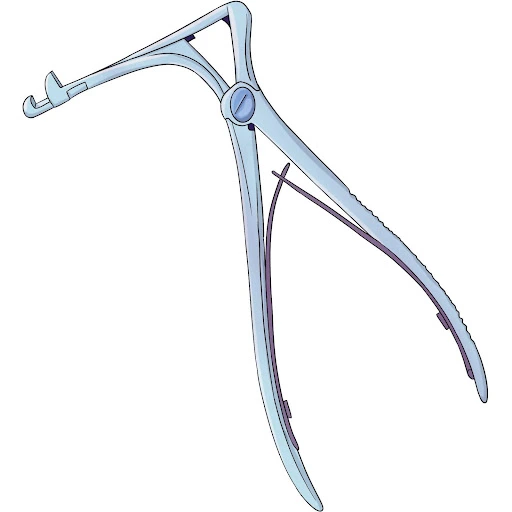
Q7. Which of the following surgeries uses this instrument?
- Evisceration
- Enucleation
- Cataract surgery
- Dacryocystorhinostomy
Ans. 4) Dacryocystorhinostomy
- The instrument shown is a, composed of a sturdy spring handle and two blades that are joined at a right angle. The upper blade features a sharp cutting edge and a small hole, while the lower blade has a cup-shaped indentation.
- This tool is utilized in DCR (dacryocystorhinostomy) surgery to expand the bony opening by punching the bone from the edges of the opening.
Also read: Last 5 Year PYQs in Anatomy for FMGE
Q8. What is the likely cause of the sudden loss of vision in the left eye for the past two hours in a 58-year-old man who experienced it after his retirement party? The fundoscopic examination reveals a similar image, as shown below.

- BRAO
- BRVO
- CRAO
- CRVO
Ans. 3) CRAO
- The given image shows a cherry-red spot. The orange reflex from the intact choroid stands out at the thin foveola, in contrast to the surrounding pale retina, giving rise to a ‘cherry-red spot’ appearance.
- Sudden loss of vision and cherry red spots are characteristic of Central Retinal Artery Occlusion.
- CRAO (Central Retinal Artery Occlusion): CRAO occurs when there is a sudden blockage in the central retinal artery, leading to a severe and immediate loss of vision. It is usually through embolism, and the risk factors include the patient being a heart patient, having carotid artery disease, or being associated with mucormycosis of the orbit.
Q9. The probable cause for a patient with a corneal scar having a visual acuity of 6/36, which improves to 6/18 with a pinhole, is most likely due to…
- Glaucoma
- Malingering
- Irregular astigmatism
- Cataract
Ans. 3) Irregular astigmatism
- The presentation suggests that the patient is a case of irregular astigmatism. Here, the refractive error of the patient improved to 6/18 using a pinhole camera, which is suggestive of refractive error.
- The history of a corneal scar that makes the surface irregular may indicate that the above case is that of irregular astigmatism.
Also read: Last 5 Year PYQs in FMT for FMGE
Q10. A male patient, aged 18, seeks medical attention due to an eye injury sustained while climbing a tree five days ago. The patient reports experiencing pain, sensitivity to light (photophobia), and redness in the affected eye for the past two days. Upon performing a slit-lamp examination, a lesion is observed as depicted below. Which of the following characteristics is associated with this particular condition?

- Reverse hypopyon
- Dendritic ulcer
- Ring abscess
- Satellite lesion
Ans. 4) Satellite lesion
- The clinical presentation and history of trauma with vegetative matter suggest that it is a case of fungal corneal ulcer. The signs are more pronounced than symptoms.
Q11. On ophthalmologic examination, the presence of Koeppe's nodules is observed in a patient diagnosed with sarcoidosis. In which area of the eye are these nodules typically observed?
- Cornea
- Conjunctiva
- Iris
- Retina
Ans. 3) Iris
- Koeppe's nodules are small, yellow-white nodules that appear on the surface of the iris and are observed in granulomatous uveitis like sarcoidosis, tuberculosis, Vogt-Kanayagi-Harada’s disease, and sympathetic ophthalmitis.
- They are typically located at the pupillary margin, which is the border between the iris and the pupil.
- These nodules are composed of collections of inflammatory cells.

Q 12. A 45-year-old male arrived at the emergency ward with sudden vision loss and experiencing painful eye movements. Upon examination, the affected eye shows a relative afferent pupillary defect, a normal optic disc, and a central scotoma. What is the likely diagnosis?
- Optic nerve glioma
- Optic neuritis
- Eale’s disease
- Optic atrophy
Ans. 2) Optic neuritis
- The sudden loss of vision, painful eye movements, relative afferent pupillary defects, and central scotoma are characteristic of optic neuritis.
- It typically presents with marked sudden vision loss, dark adaptation may be lowered, and decreased contrast sensitivity.
- On examination, RAPD (relative afferent pupillary defect), central scotoma, and centrocecal scotoma can be seen.
Also read: Last 5 Year PYQs in OBG for FMGE
Q13. When selecting the strength of an intraocular lens (IOL) for a child under 1 year old, what factors should be considered?
- Myopic shift
- Hypermetropic shift
- Length of the lens capsule
- Growth of the lens capsule
Ans. 1) Myopic shift
- When choosing the power of an intraocular lens (IOL) in a child under 1 year of age, the myopic shift should be taken into account.
- A myopic shift is a phenomenon where the eye becomes more myopic as it grows, and this can lead to a child becoming increasingly nearsighted over time.
- To compensate for this, a lower power IOL should be chosen to allow for the myopic shift that will occur as the child's eye grows.
- The length of the lens capsule and growth of the lens capsule are important factors to consider when selecting an IOL, but they are not the primary factors in choosing the power of the IOL in a young child.
Q14. Please analyze the image provided and determine the specific abnormality or injury present.

- Viral keratitis
- Fungal keratitis
- Bacterial keratitis
- Interstitial keratitis
Ans. 1) Viral keratitis
- The lesion in viral keratitis is typically characterized by dendritic or branching patterns on the cornea, as seen in the image.
- Viral keratitis is a type of corneal infection caused by a virus, such as herpes simplex virus (HSV) or varicella-zoster virus (VZV).
Also read: Last 5 Year PYQs in Anesthesia for FMGE
Q15. What is the likely reason for a 21-year-old student to have ocular redness, feeling of a foreign object in the eye, and excessive tearing after wearing a soft contact lens, as indicated by the corneal scraping interface contrast microscopy image?

- Acanthamoeba
- Naegleria fowleri
- Entamoeba histolytica
- Balamuthia mandrillaris
Ans. 1) Acanthamoeba
- The clinical presentation of ocular redness, foreign body sensation, epiphora, history of using a soft contact lens, and image showing a cyst of acanthamoeba characterized by an outer wrinkled wall is suggestive of acanthamoeba keratitis caused by acanthamoeba, a free-living protozoan.
- It is prevalent in individuals using soft contact lenses and using tap water for cleaning or swimming with the contact lenses in a swimming pool.
Hope you found this blog helpful for your FMGE Medicine Preparation. For more informative and interesting posts like these, keep reading PrepLadder’s blogs.
Download PrepLadder's FMG Exam preparation app for Android
Download PrepLadder's FMG Exam preparation app for iOS
Also check the Previous Year Question Papers for FMG Exams.
.jpg)

PrepLadder Medical
Get access to all the essential resources required to ace your medical exam Preparation. Stay updated with the latest news and developments in the medical exam, improve your Medical Exam preparation, and turn your dreams into a reality!
Top searching words
The most popular search terms used by aspirants
- FMGE Ophthalmology
- FMGE Ophthalmology Perparation
PrepLadder Version X for FMGE
Avail 24-Hr Free Trial