Last 5 Year PYQs in Biochemistry for FMGE
Jan 27, 2025

When it comes to preparing for FMGE, there is no smarter way to understand exam patterns, identify high-yield topics, and refine your strategy than solving previous years’ questions (PYQs).
As Biochemistry is a volatile and high-scoring subject, it requires conceptual clarity and consistent revision to ace the exam.
But, what if you had access to the most frequently asked questions from the last five years. That’s exactly what we’ve got for you in this blog.
We have compiled high-yield Biochemistry PYQs that have been frequently tested in FMGE. And we have included detailed explanations to help you understand the concepts better nad improve your retention.
Without further ado, let’s dive right in.
Q1. On centrifugation of a cell, LDH will get deposited in:
- Mitochondria
- Lysosome
- Cytoplasm
- Golgi complex
Answer 3) Cytoplasm
- LDH, commonly known as lactate dehydrogenase, is a protein that functions as an enzyme. It is a tetrameric enzyme consisting of two monomer types: H (for the heart) and M (for muscle) that combine to yield five LDH isozymes: HHHH (I1), HHHM (I2), b, HMMM (I4), and MMMM (I5). The enzyme of glycolysis, which takes place in the cytoplasm, is lactate dehydrogenase. It serves as a cytoplasmic marker.
Q2. The biochemical form of amyloid in hemodialysis-associated amyloidosis is:
- AL
- AA
- Aβ2m
- ATTR
Ans. 3) Aβ2m
- Amyloidosis is a protein-folding disorder in which normally complete proteins or fragments of normally soluble proteins accumulate as aberrant, insoluble fibrils that damage tissue structure and cause disease.
- Aβ2M is a small membrane protein associated with the heavy chains of class I major histocompatibility complex (MHC) on the surface of nucleated cells. Most of the β2M is typically cleared by glomerular filtration and subsequent proximal tubular reabsorption and catabolism. An increase in the concentration of β2-macroglobulin (β2m) in the blood appears to be a requirement for Aβ2M amyloidosis, which results in Aβ2M amyloid deposition primarily in the osteoarticular tissue.
Also read: FMGE January 2025 Admit Card Out: Download Yours Now!
Q3. A 13-year-old with acute flaccid paralysis, the pediatric resident orders an RT-PCR on a CSF sample to identify the causative virus. Which of the following described the enzyme correctly?
- DNA-dependent RNA polymerase
- RNA-dependent DNA polymerase
- DNA-dependent DNA polymerase
- RNA-dependent RNA polymerase
Ans. 4) RNA-dependent RNA polymerase
- Poliovirus is most commonly recovered from stool specimens.If poliovirus is isolated, the serotype (i.e., 1, 2, or 3) and whether the virus is wild, vaccine (Sabin), or VDPV strain are determined using reverse transcriptase polymerase chain reaction (RT-PCR) and genomic sequencing.
- Because acute flaccid paralysis (AFP) mirrors the clinical look of poliomyelitis, it has been accepted as a critical tool for tracking the global polio eradication effort's success.
- If poliovirus is isolated, the serotype (i.e., 1, 2, or 3) and whether the virus is wild, vaccine (Sabin), or VDPV strain are determined using reverse transcriptase polymerase chain reaction (RT-PCR) and genomic sequencing.
- In ssRNA viruses, RNA genome replication occurs in a membrane-bound replication complex composed of the viral polymerase and other viral proteins, cellular proteins, and viral RNA. The replicating complex's specific content and nature currently need to be clarified. Plus-sense genomic RNA functions as an mRNA in infected cells to make viral proteins without modifying the viral genome, and hence viral RNA-dependent RNA polymerases are responsible for viral RNA genome replication.
Q4. Which one of the following is not classified as a micromineral?
- Calcium
- Selenium
- Iodine
- Copper
Ans. 1) Calcium
- Calcium is considered a macromineral because it is required by the body in larger amounts compared to microminerals. Macrominerals are needed in quantities greater than 100 milligrams per day.
- Minerals are divided into two types: macrominerals and microminerals.
- Macrominerals, including calcium, phosphorus, magnesium, are required in amounts larger than 100 mg daily.
- Microminerals, which include electrolytes including sodium, potassium, chloride, iron, copper, zinc, selenium, and iodine, are required at levels less than 100 mg per day.
Also read: The Ultimate 5-Day Strategy to Ace FMGE Jan ‘25
Q5. What is the likely diagnosis for a 36-year-old man who has been experiencing bluish-black discoloration of the sclera and pinna for the past 6 months, along with a history of black urine upon standing since childhood?
- Phenylketonuria
- Alkaptonuria
- Tyrosinemia
- Maple syrup urine disease
Ans. 2) Alkaptonuria
- Alkaptonuria is an uncommon hereditary condition. It occurs when the body cannot make enough homogentisic dioxygenase (HGD). This enzyme is utilized to degrade the hazardous chemical homogentisic acid. When the body does not make enough HGD, homogentisic acid accumulates. The accumulation of homogentisic acid causes discoloration and brittleness of bones and cartilage. This usually results in osteoarthritis, particularly in your spine and big joints. When exposed to air, in those with alkaptonuria urine turns dark brown or black.
Q6. Complex IV is inhibited by--?
- Rotenone
- Cyanide
- Carboxin
- Antimycin A
Ans. 2) Cyanide
- The Electron Transport Chain (ETC) is a collection of four protein complexes (NADH dehydrogenase (Complex I), succinate dehydrogenase (Complex II), cytochrome b and c1 (Complex III), and cytochrome c oxidase (Complex IV)) that couple redox processes, resulting in an electrochemical gradient that leads to the formation of ATP in a process known as oxidative phosphorylation. It happens in both cellular respiration and photosynthesis in mitochondria. In the former, electrons are released as a result of the breakdown of organic molecules. In the latter, electrons are stimulated by light and join the chain, where the energy released is used to construct carbs.
- The suppression of cytochrome c oxidase (CcOX), the oxygen-reducing component of mitochondrial electron transport, is thought to be the cause of acute cyanide toxicity.
- Cyanide is regarded as a cytotoxic agent in mammalian cells, where it acts by inhibiting mitochondrial Complex IV (Cytochrome C oxidase [CCOx]). However, the current study shows that cyanide has a biphasic effect on CCOx: low (nanomolar to low-micromolar) concentrations increase CCOx activity, while higher (high-micromolar) concentrations have the "classic" inhibitory effect. Cyanide at low concentrations promoted mitochondrial electron transport and increased intracellular adenosine triphosphate (ATP), stimulating cell growth.
Also read: Don’t Panic! The Perfect Last-Minute FMGE Strategy
Q7. Which of the subsequent stages in the vitamin D metabolism necessitates exposure to sunlight?
- 1,25 dihydroxy cholecalciferol to 7-dehydrocholesterol
- 7-dehydrocholesterol to cholecalciferol
- Cholecalciferol to 25-hydroxycholecalciferol
- 25-hydroxycholecalciferol to 1,25 dihydroxycholecalciferol
Ans. 2) 7-dehydrocholesterol to cholecalciferol
- Vitamin D is produced endogenously in the body in a process that involves the skin, liver and kidneys.
- 7-dehydrocholesterol is present in the epidermis of the skin. The UVB radiation present in sunlight reacts with 7-dehydrocholesterol converting it into cholecalciferol. Sunlight plays a crucial role in this conversion without which the Vitamin D synthesis does not progress further.
Q8. What is the cause of Menke's disease?
- Zinc
- Copper
- Iron
- Magnesium
Ans. 2) Copper
- Menke’s disease occurs due to a deficiency of copper due to a defect in the absorption of copper from the intestine. The copper gets trapped in the metallothionine present in the intestinal cells. This prevents the copper from being absorbed into the bloodstream. The symptoms of Menke’s disease include reduced copper in plasma and urine, anemia and depigmentation of hair.
Q9. Which enzyme is responsible for the deficiency causing classical galactosemia?
- Aldose reductase
- Galactokinase
- Galactose-1-phosphate uridyl transferase
- UDP galactose-4-epimerase
Ans. 3) Galactose-1-phosphate uridyl transferase
- Classical Galactosemia is due to the deficiency of the enzyme Galactose-1-phosphate uridyl transferase (GALPUT).
- It is a rare congenital autosomal recessive disorder. In this condition, galactose levels are increased in circulation and urine. The accumulated galactose-1-phosphate is converted into dulcitol by the enzyme aldose reductase. This accumulation of Dulcitol is associated with the development of cataracts., and impairment of organs such as the liver and kidney.
Also read: Biochemistry Important Questions For NEET PG/FMG Exams
Q10. Which one of the following choices does not represent a function of glucagon?
- Glycolysis
- Gluconeogenesis
- Glycogenolysis
- Ketogenesis
Ans. 1) Glycolysis
- Glucagon is a hormone secreted by the alpha cells of islets of Langerhans in the pancreas.
- Glucagon does not play a role in Glycolysis which is the process of breakdown of glucose as it reduces blood glucose levels.
- On the contrary, glucagon increases the synthesis of glucose (gluconeogenesis) and the breakdown of glycogen in the liver to release glucose (Glycogenolysis). The levels of Glucagon in blood increase in fasting state.
Q11. What could be the potential diagnosis for a 34-year-old male who has been consuming maize as his main diet for an extended period and is now experiencing symptoms such as abdominal pain, decreased appetite, and diarrhea? Additionally, upon examination, glossitis (inflammation of the tongue) and a pigmented rash in sun-exposed areas were observed, along with a lesion on his neck?
- Pellagra
- Scurvy
- Megaloblastic anemia
- Beriberi
Ans. 1) Pellagra
- Pellagra is a condition caused by the deficiency of Niacin ( Vitamin B3) in the body. The symptoms seen in the patient including pain in the abdomen, loss of appetite and diarrhoea along with glossitis and pigmented rash in sun-exposed areas are symptoms commonly associated with Pellagra.
- Since the patient has a staple diet of Maize which does not contain Vitamin B3 and does not have any other source of niacin, it is conclusive that the diagnosis, in this case, is Pellagra.
Also read: Important Questions On Carbohydrate Chemistry
Q12. Due to a prolonged period of consuming maize as a main food source, a 34-year-old man developed diarrhoea and exhibited lesions on his neck, as depicted in the accompanying image. This condition is caused by a deficiency in:

- Niacin
- Zinc
- Thiamine
- Iron
Ans. 1) Niacin
- The above picture presented above suggests Vitamin B3/niacin deficiency according to clinical features. Pellagra disease is caused by the excessive consumption of a diet made up of maize. Maize/corn has low levels of tryptophan and niacin.
- Niacin deficiency causes dermatitis, diarrhoea, and dementia.
- The neck lesion shows characteristic Casal’s necklace pattern, as seen in Pellagra. The Casal necklace or Casal collar is a clinical sign of pellagra that shows erythematous pigmented rash of skin near the collarbone region (dermatomes C3 and C4).
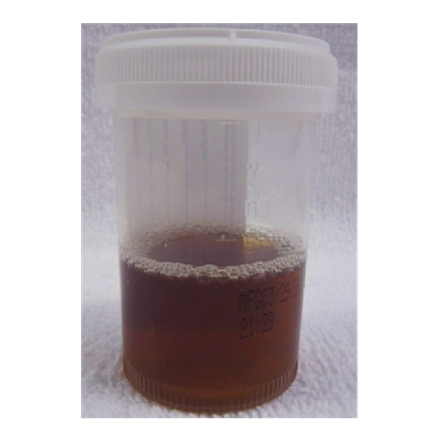
Q13. The lab technician observes that a urine sample obtained from a 4-year-old male appeared normal initially, but underwent a change in color as depicted in the image after a few hours. Subsequently, the color intensified over the following hours. Which deficiency can potentially lead to this condition?
- Beta-carotene
- Tyrosine aminotransferase
- L-ascorbic acid
- Homogentisic acid oxidase
Ans. 4) Homogentisic acid oxidase
- Homogentisic acid (HGA) is a metabolite that is used as the diagnostic substance in the tissues for alkaptonuria. Alkaptonuria is an autosomal recessive disease that occurs rarely.
- When Homogentisic-acid oxidase is found deficient in the tissue, then normal procedure of phenylalanine metabolism gets blocked and Homogentisic acid (HGA) becomes unable to break. Hence, urine appears as black and dark-stained nappies, and can be seen as the earliest signs of it.
Q14. Complex IV is inhibited by?
- Rotenone
- Cyanide
- Carboxin
- Antimycin A
Ans. 2) Cyanide
- Cyanide compound poisons the electron transport chain in the mitochondria within the cells. It stops the body from deriving energy (ATP) from oxygen. Specifically, cyanide attaches with the a3 prosthetic group in the complex IV of cytochrome C oxidase (CCOx) and causes death of the cell rapidly. The generation of ATP is shutdown in the aerobic respiration that is recognized as cyanide toxic actions in the eukaryotes primarily.
Also read: Most IMP Biochemistry PYQs for FMGE July ‘23
Q15. Which of the statements below accurately describes the characteristics of the Michaelis-Menten graph?
- Km is directly proportional to substrate concentration
- Km doesn't vary with the enzyme concentration
- Km is indirectly proportional to substrate concentration
- Km has no relation to substrate concentration
Ans. 1) Km is directly proportional to substrate concentration
- Km stands for the dissociation constant of enzymes. A high Km represents weak binding of enzymes with the substrate while a low Km stands for strong binding with the substrate. Hence, dissociation of substrate does not occur if a low Km value is present.
- According to Michaelis-Menten equation,
- v = Vmax [S] /Km+[S]
- where v is the velocity of enzyme, S is substrate, Km is Michaelis-Menten constant, and Vmax is maximum velocity achieved by the enzyme. Hence, the rate of enzyme and substrate concentration is directly proportional to each-other. The reaction belongs to first-order kinetics.
Q16. By the action of the enzyme aromatase, testosterone is transformed into which of the subsequent hormones?
- 5-hydroxy testosterone
- Estradiol
- Cortisol
- Cortisone
Ans. 2) Estradiol
- Aromatase enzyme is used in the process of androgen (such as testosterone) to estrogen (such as 17β-estradiol) conversion. Aromatase enzyme is used as a therapeutic target for the treatment process of endocrine-responsive breast cancer. Aromatase also converts androstenedione to estrone.
Q17. A farmer sprayed a chemical in the pool which causes inhibition of complex-III of the electron transport chain. Identify the inhibitor?
- Rotenone
- TTFA
- Cyanide
- Antimycin
Ans. 4) Antimycin
- Antimycin is an inhibitor of complex III (cytochrome b-c1 complex) of the electron transport chain. It interrupts the transfer of electrons from ubiquinol to cytochrome c, thereby inhibiting the flow of electrons through this complex.
Also read: Fatty Acid Oxidation - NEET PG Biochemistry
Q18. Cholera is known to cause diarrhea through its interaction with specific receptors in the human body. Which of the following is known to make it easier for cholera toxin to enter the body through the intestinal mucosa?
- GLUT 1
- GM2 ganglioside
- Cephalin
- GM1 ganglioside
Ans. 4) GM1 ganglioside
- Cholera toxin is produced by the bacterium Vibrio cholerae and is responsible for causing the characteristic symptoms of cholera, including severe diarrhea. Cholera toxin enters the body through the intestinal mucosa and interacts with specific receptors on the surface of intestinal cells.
- The receptor that facilitates the entry of cholera toxin into the body is the GM1 ganglioside receptor. GM1 ganglioside is a glycosphingolipid, a type of molecule found in the lipid bilayer of cell membranes. Cholera toxin binds to GM1 ganglioside on the surface of intestinal cells, which triggers the toxin to be internalized into the cell through endocytosis. Once inside the cell, the toxin can exert its effects, leading to the disruption of ion transport and fluid balance, ultimately causing severe diarrhea.
Q19. A 2-month-old infant is brought to the pediatrician due to feeding difficulties, vomiting, and failure to thrive. The child's history reveals that the infant was born full-term and had a normal birth weight. On physical examination, the infant appears jaundiced, and the liver is palpable below the costal margin. Oil drop Cataracts are also noted on eye examination. Laboratory tests show elevated levels of galactose in the blood and urine. Based on these clinical findings, which enzyme deficiency is most likely responsible for this presentation?
- G6PD (Glucose-6-phosphate dehydrogenase)
- GALT (Galactose-1-phosphate uridylyltransferase)
- ALDH (Aldehyde dehydrogenase)
- PKU (Phenylalanine hydroxylase)
Ans. 2 - GALT (Galactose-1-phosphate uridylyltransferase) deficiency
- GALT deficiency, also known as galactosemia, is a genetic disorder that impairs the ability to metabolize galactose properly. Galactose is a sugar found in milk and other dairy products.
- In individuals with GALT deficiency, galactose-1-phosphate accumulates, leading to various symptoms, including feeding difficulties, vomiting, failure to thrive, jaundice, and liver enlargement.
- Cataracts are also a characteristic feature of galactosemia. This condition requires the removal of galactose from the diet to prevent complications.
Also read: Lipids: Classification, Pufas, Ketone Body Synthesis
Q20. A newborn infant is brought to the clinic with jaundice and a white, punctate, and scattered opacity in the central lens. The condition is consistent with Oil drop cataract. Which of the following metabolic disorders is most likely responsible for this presentation?
- Phenylketonuria (PKU)
- Maple syrup urine disease
- Galactosemia
- Wilson's disease
Ans. 3) Galactosemia
- The most likely metabolic disorder responsible for the presentation of oil drop cataract in a newborn infant with jaundice and lens opacities is Galactosemia. Oil drop cataract is a type of cataract characterized by small, white, punctate opacities that resemble oil drops in the lens of the eye. This specific type of cataract is associated with galactosemia, which is an inherited metabolic disorder that affects the body's ability to metabolize galactose, a sugar found in milk and other dairy products.
- In galactosemia, there is a deficiency in one of the enzymes involved in the metabolism of galactose, leading to the accumulation of galactose-1-phosphate in various tissues, including the lens of the eye. The accumulation of galactose-1-phosphate can result in the formation of the characteristic oil drop opacities seen in the lens.
Hope you found this blog helpful for your Best FMGE PG Coaching online. For more informative and interesting posts like these, keep reading PrepLadder’s blogs.
Download PrepLadder's best app for fmge preparation for Android
Download PrepLadder's best app for fmge preparation for Ios


PrepLadder Medical
Get access to all the essential resources required to ace your medical exam Preparation. Stay updated with the latest news and developments in the medical exam, improve your Medical Exam preparation, and turn your dreams into a reality!
Top searching words
The most popular search terms used by aspirants
- FMGE Biochemistry
- FMGE Biochemistry Preparation
PrepLadder Version X for FMGE
Avail 24-Hr Free Trial